
.jpeg?width=75&height=75&name=Carlos%20Ros%20(Default%20-%20Team%20Sheet).jpeg)

This article explores Ischemic myelopathy (IM), a vascular spinal cord condition in small animals, commonly caused by fibrocartilaginous embolism (FCE). It discusses IM etiology, clinical signs (like per-acute, non-painful, non-progressive neurological dysfunction), diagnostic pathways (including MRI findings), and management options with prognosis based on severity and imaging.
Ischemic myelopathy (IM) is a vascular condition of the spinal cord that results from sudden interruption of blood flow, often due to embolic obstruction. It is a relatively common clinical situation in small animals and can lead to varying degrees of spinal cord ischemia and, in some cases, necrosis of the spinal cord parenchyma. However, not all cases result in necrosis; the severity depends on the extent and duration of the ischemia.

Etiology of ischemic myelopathy
The most common described cause of spinal blood vessel obstruction are fibrocartilaginous emboli, and for that reason, this condition is called “fibrocartilaginous embolism” (FCE). This fibrocartilaginous material is histologically and histochemically identical to the nucleus pulposus of the intervertebral disc.
Although the most common cause of IM is FCE, other causes including thromboembolism, hypercoagulability, vasculopathy, and septic, parasitic, neoplastic and fat embolization have also been reported. IM can also happen secondary to a vascular spasm after a traumatic event. In cats, IM is generally secondary to an underlying predisposing condition such as systemic hypertension, chronic kidney disease, cardiomyopathy, hyperthyroidism and hyperaldosteronism.
Features of small animal ischemic myelopathy
Dogs: It is reported most commonly in large and giant breed dogs of non-chondrodystrophic breeds. However, it can also happen in small breed dogs, particularly in miniature Schnauzers. The typical median age is 4-6 years (range 2 months to 13 years) (Theobald A et al, 2013).
Cats: The most represented breed is the Domestic Short Hair. The typical median age is 10 years (range 6 months – 17 years) (Theobald A et al, 2013)
Typical clinical presentation:
- Per-acute onset (<6 hours) of neurological dysfunction (referable to the site and extent of the spinal cord injury)
- Non-painful condition (after the first 24–36 hours)
- Non-progressive disease (after the first 24-36 hours)
Lesion location:
- Dogs: There are discrepancies between different studies regarding the most common location in the spinal cord, and therefore, FCE can occur in any spinal cord segment.
- Cats: FCE has been reported to occur more frequently in the cervical spinal cord than other locations, as the ventral spinal artery is narrowest at C2 vertebra.
Differential diagnoses and diagnostic pathways for small animal ischemic myelopathy
Differential diagnosis:
- Other types of intervertebral disc disease: Acute non-compressive hydrated nucleus pulposus extrusion and intervertebral disc extrusion of a degenerated intervertebral disc
- Intramedullary neoplasia
- Infectious and non-infectious meningomyelitis
Diagnostic approach:
The definitive diagnosis of IM can only be obtained by histopathological examination, however, the antemortem diagnosis can be suspected and reached based on the typical clinical presentation and exclusion of other causes of similar presentation.
As in any neurological patient, hematology, complete biochemistry, urinalysis, measurement of blood pressure, thoracic radiographs and abdominal ultrasound may be indicated before performing advanced imaging techniques (e.g magnetic resonance imaging (MRI)).
In feline patients, measurement of T4 levels and cardiac ultrasound is also indicated.
Magnetic resonance imaging findings:
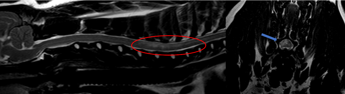
The lesion is typically a focal, well-defined and sharply demarcated intramedullary, and often lateralized, lesion, predominantly involving the gray matter. The lesion is generally hyperintense in T2-weighted and FLAIR images, and iso- or hypointense to normal gray matter on T1-weighted images. On occasion, MRI can be normal (if performed 24-72 hours after onset). Contrast enhancement can be observed generally five to seven days after the onset of clinical signs. Generally, the intramedullary lesion is overlying a vertebral body (not over the intervertebral disc space).
Recently, the use of reduced field of view diffusion-weighted imaging MRI has been reported in dogs and cats with suspected acute ischemic myelopathy. These patients showed restricted diffusion, consistent with ischemia and cytotoxic edema.
Other imaging modalities to use for ischemic myelopathy
Myelography
Myelography can be normal or may reveal an intramedullary pattern suggesting spinal cord swelling in the acute stage of the disease. Myelography helps to rule out other causes of acute myelopathy.
Computed tomography
CT also helps to rule out other causes of acute myelopathy. An intramedullary pattern can also be observed on myelo-CT.
Cerebrospinal fluid analysis
CSF analysis is a very sensitive but non-specific diagnostic technique. In patients with IM/FCE, CSF analysis can be normal or may reveal non-specific abnormalities. i.e: xanthochromia, high protein content, and mild to marked pleocytosis.
Management and prognosis of ischemic myelopathy
Management: Treatment is conservative. Anti-inflammatory (NSAIDs) and analgesic medications are indicated in patients with spinal hyperesthesia. If IM has occurred secondary to trauma, further assessment for concurrent injuries should be addressed. Fluid therapy may be indicated to guarantee spinal cord perfusion. It is important to make sure that blood pressure, oxygenation, and ventilation are within normal values. Nursing care, rest and physiotherapy (if there are significant motor deficits) are also recommended.
Outcome and prognosis: The prognosis depends on the degree of tissue damage. Loss of nociception has been reported as a negative prognostic factor. Other negative prognostic factors are lower motor neuron signs and symmetrical neurological deficits.
MRI can also help provide prognostic information. Certain features seen on imaging, such as the length or size of the spinal cord lesion, have been associated with poorer outcomes. For example, larger or more extensive changes on MRI may indicate a less favourable prognosis.
References
De Risio L. A review of fibrocartilaginous embolic myelopathy and different types of peracute non-compressive intervertebral disk extrusions in dogs and cats. Front Vet Sci. 2015 Aug 18;2:24. doi: 10.3389/fvets.2015.00024. PMCID: PMC4672181
Theobald A, Volk HA, Dennis R, et al. Clinical outcome in 19 cats with clinical and magnetic resonance imaging diagnosis of ischaemic myelopathy (2000-2011). J Feline Med Surg. 2013 Feb;15(2):132-4. doi: 10.1177/1098612X12463927. PMID: 23048075
De Risio L, Adams V, Dennis R, McConnell FJ, Platt SR.Association of clinical and magnetic resonance imaging findings with outcome in dogs suspected to have ischemic myelopathy: 50 cases (2000-2006). J Am Vet Med Assoc. 2008 Jul 1;233(1):129-35. doi: 10.2460/javma.233.1.129. PMID: 18593322
Explore more

Jun 5, 2025 12:53:38 PM
Neutropenia - a haematologic adverse event of chemotherapy
This article considers bone marrow suppression, an important chemotherapy side effect. It details...
Read more
Jun 5, 2025 12:53:38 PM
Diagnostic Approach to Hypercalcaemia in dogs
This guide details the diagnostic approach to hypercalcaemia in dogs, covering clinical signs...
Read more