

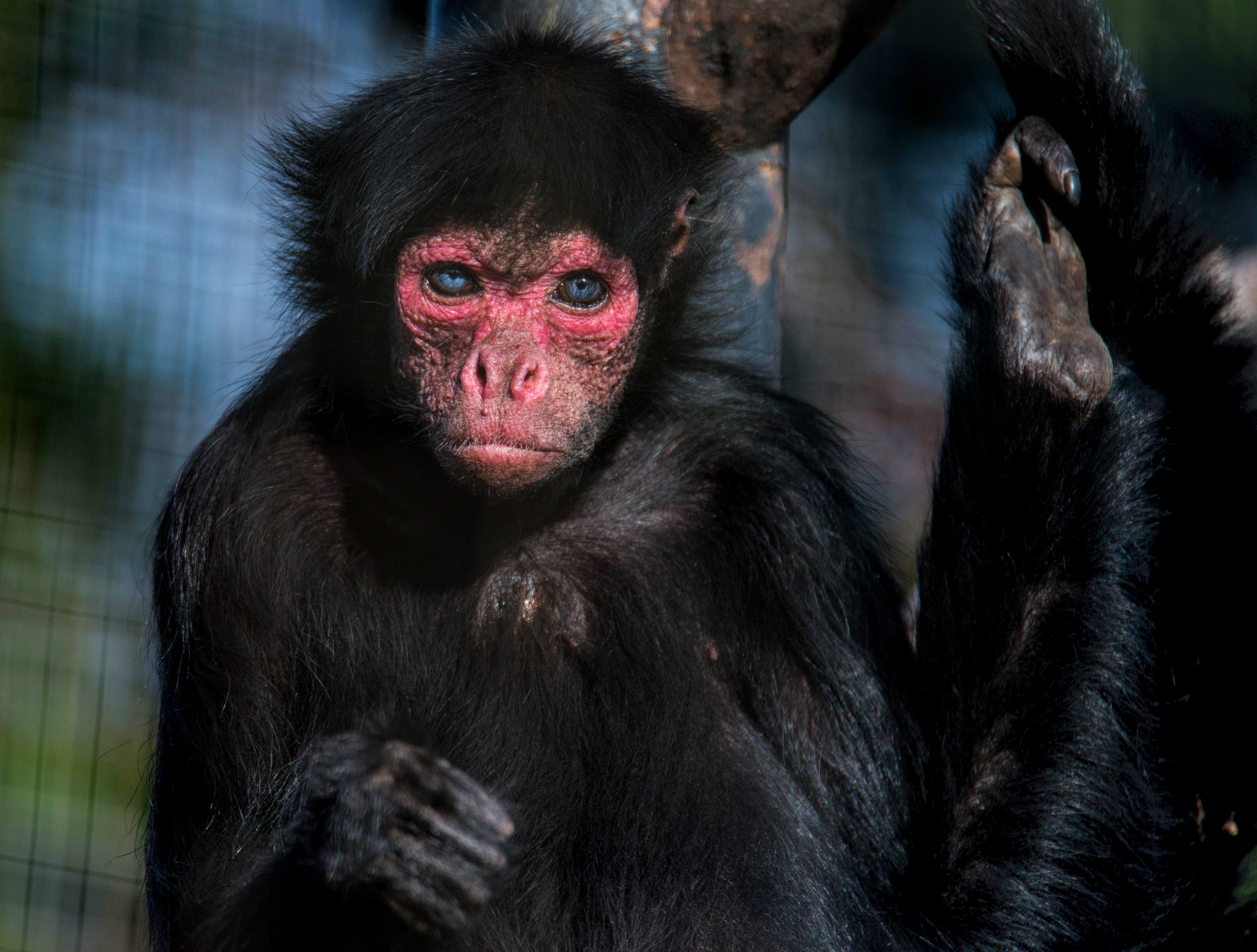
Smokey, a rare red-faced spider monkey from the Welsh Mountain Zoo in Colwyn
Bay, Conwy had become unwell and started vomiting. The zoo had lost a female
spider monkey a few months prior, due to a perforated ulcer.
Keepers acted quickly, and had Smokey examined by the team at Mochdre Vets,
who recommended a CT scan to diagnose the problem. While Smokey was under anaesthetic, the images were sent to a remote diagnostic imaging specialist from VET.CT for immediate reporting.
Maya Esmans, located in Belgium, read the images and conferred with VET.CT colleagues to confirm the most likely diagnosis as gastroenteritis.
Deputy Head Nurse Robi McCaffrey said, "The scan meant we could treat Smokey
without surgery. We were able to rule out tumours, foreign bodies, and even ulcers.”
VET.CT Report Findings:
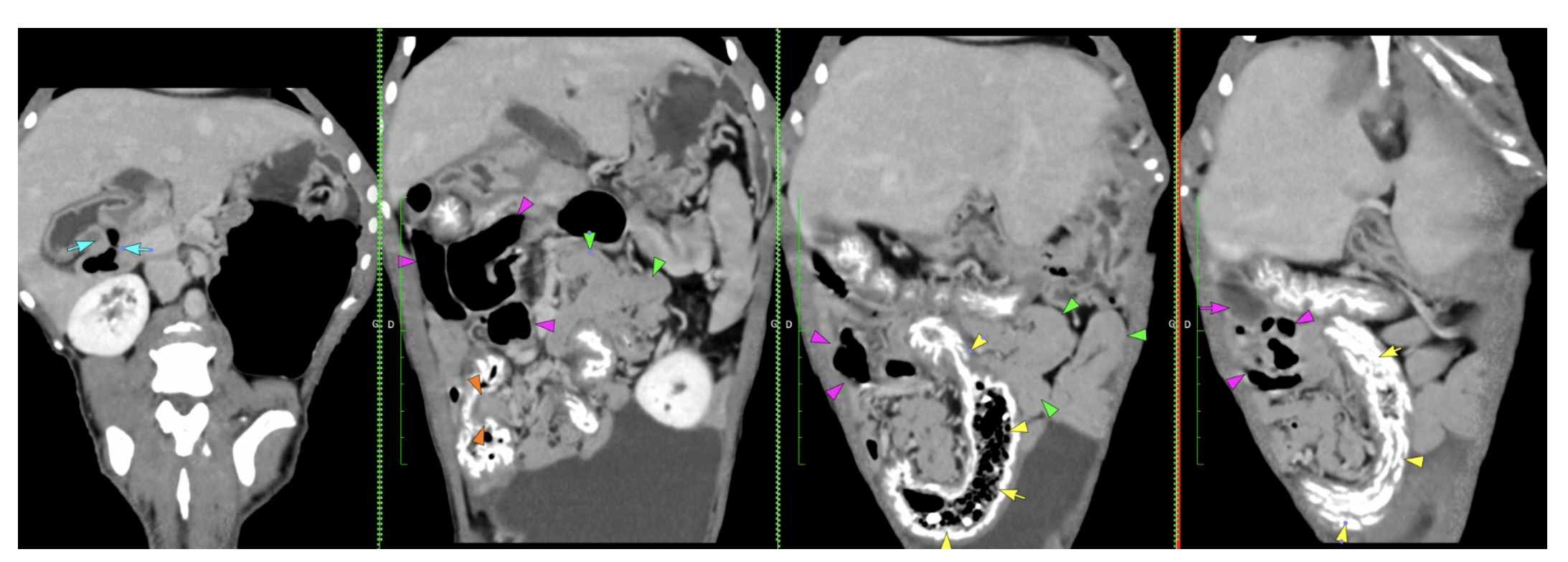
The stomach is moderately distended with gas and a small amount of fluid. The pylorus is well defined. Some gas bubbles are seen just after the pylorus (blue arrows), then the duodenum is rather poorly filled and difficult to completely follow.
A small to medium amount of gas and fluid is seen accumulating in the jejunum in the right mid-abdomen (pink arrows), but it seems to get to a normal size on both sides without evidence of luminal obstructive foreign material. The rest of the jejunum is rather prominent but almost empty (green arrows).
The mesenteric lymph nodes are difficult to delineate and measure precisely, but do not seem significantly enlarged.
The ileocaecal junction is well seen (orange arrows). Mineral coating is seen all along the caecum and colon (previous barium study versus mineral oral coating).
Some gas-mixed feces are seen in the caecum (yellow arrows), the colon is almost empty, partially multifocally gas-distended.
I discussed this case with my colleague, as there is a doubtful region in the right mid-abdomen. We cannot delineate luminal foreign material, and the distension remains moderate while the rest of the gastrointestinal tract is rather empty. We suspect gastroenteritis given the absence of a clear obstruction. An infiltrative pathology with mild thickening is also possible, but it would not fit the acute clinical signs.
Reported by: Dr Maya Esmans, DMV, MRCVS, ECVDI European Specialist in Veterinary Diagnostic Imaging
Head nurse Jess Nettleton said: "The procedure is very quick, which meant Smokey
was only asleep for a short time - something that carries obvious benefits for his
safety and recovery. By the same afternoon, he was safely back in his pen and
eating again."
Zoo staff say Smokey has now recovered and is now eating and acting as normal
following a course of treatment. Commercial Director at the Welsh Mountain Zoo Michaella Brannan said "Smokey is much better and is back to his normal self, interacting with visitors. He's now back to eating a normal diet and swinging effortlessly around his enclosure in the way that we'd expect him to."
Remote diagnostic imaging reporting is invaluable in providing expertise at the point
of care for patients. VET.CT’s team of over 150 board-certified radiologists are
located all over the world, providing 24/7 radiology reports and providing imaging
advice and reports for all species to veterinary teams worldwide, supporting them
with accurate, timely diagnostic reports to inform patient care.

Explore more
-2.png)
Oct 23, 2025 9:30:00 AM
Video Guides to Veterinary CT Scanning - Head and Neck
Video guides to veterinary CT scanning - head and neck Computed Tomography (CT) is a useful...
Read more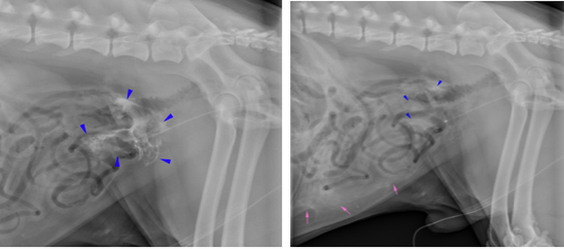
Oct 23, 2025 9:30:00 AM
It's Your Case - 2yo Labrador Dog with abdominal pain after vehicular trauma.
Welcome to It's Your Case! In the next instalment of It's Your Case, we take a look at a case of a...
Read more