

The process of teleradiology involves two critical components: the expert interpretation of the medical images and the effective communication of those findings in a clinically useful, comprehensive report. Mastering both is essential for success, but most importantly of all, we really care about the patients we serve and the teams looking after them. At VET.CT, our core mission is to provide clear, accurate, and actionable diagnostic imaging interpretations that empower our referring veterinary surgeons to deliver great care to their patients.
The VET.CT approach to diagnostic imaging reporting builds on the foundation of the 6 C's of good radiology reporting, originally attributed to R.R. Armas1:
- Clarity
- Correct
- Confidence level
- Concise
- Complete
- Consistent
To this we add two additional key components:
- Communication
- Care

VET.CT applies these principles to our expert veterinary diagnostic imaging interpretations in the following ways:
Clarity:
VET.CT reports must be unambiguous and easy for the referring veterinarian to understand. Our 150+ radiologists strive to paint a clear picture of the findings through both words and annotated images.
Correct:
Accuracy is paramount. Backed by our unique clinical warranty, VET.CT strives to provide thorough representations of relevant imaging findings and provide prioritized differential diagnoses that are well-supported by the evidence presented in the study and case history. Our commitment to quality assurance with internal second opinions provided by world-leading radiologists ensures that our interpretations are accurate, building trust and helping to avoid errors that could negatively impact patient care.
Confidence Level:
Our reports reflect the radiologist’s level of certainty. Where a definitive diagnosis can be made, we use direct, confident language. When the findings are non-specific, we provide a list of relevant differential diagnoses and recommendations for further investigation, such as laboratory tests or follow-up imaging, guiding the referring veterinarian toward the next logical steps without overstating conclusions.
Concise:
While comprehensive, the report must be succinct. We follow a clear, logical structure to facilitate rapid understanding and retrieval of key information:
- Patient and Study Details: Signalment, accession number, and the date and time of the study.
- Clinical History: A brief summary of the patient's clinical signs, relevant history, and any key questions from the referring vet.
A typical summary history from a VET.CT report:
Clinical History:
4 month old male Tonkinese fell off bed this morning and became acutely non-weight bearing.
Questions to be answered:
RHL: confirm slipped capital physis and no other abnormalities - as possible, given stool in colon
LHL: distal to stifle, possible bony changes to tibia and fibula, best seen as a possible step of one region of cortical bone on the frog-legged VD; differentials or thoughts?
- Imaging Technique: Details on the modalities and views acquired.
- Detailed Findings: A structured description of all salient findings, organised by body system.
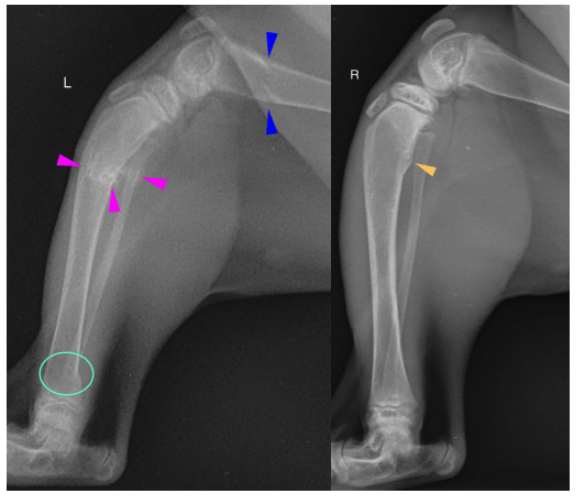
Diagnostic interpretation:
The femoral cortices are thin as compared to the tibial cortices. There are oblique folding fractures in the left distal femoral metaphysis (dark blue arrowheads) and left tibial and fibular proximal diaphyses (light pink arrowheads). Faint periosteal proliferation surrounds the fracture margins of the tibial and fibular fracture sites. The distal femoral segment is mildly cranially angulated with cranial angulation of the proximal tibial segment. There is a stair-step like lesion in the left tibial distal diaphyses (light mint circle) in the caudal cortex, as compared to the right.
The right tibial proximal metaphyseal caudal cortex is bulging caudally with smooth increased periosteal proliferation (tan arrowhead).
The left talar ridges are sclerotic and the margins are ill-defined from the distal tibial epiphysis on the lateral image, as compared to the right. The right and left calcanei are lucent within the body, with an ill-defined trabecular pattern and the cortices are thin.
The right hind soft tissues are thicker than the left cranially, however, the right limb is more flexed than the left.

VD image. There is mild increased sclerosis of the right femoral neck as compared to the left.
Additionally, there is faint, thin osseous formation along the cranial margin of the right femoral neck (white arrowhead). On the lateral image, the right proximal physis is indistinct. Increased soft tissue opacity surrounds the left coxofemoral joint.

- Conclusions: The most important section, summarising the primary findings, answering key questions, providing a likely diagnosis or differential list, and ranking the most probable causes.
• Left femoral distal metaphyseal and left tibia/fibular proximal diaphyseal pathological folding fractures
• Wide left proximal femoral physes, likely indicates a capital physeal fracture, with regional soft tissue swelling
• Right tibial proximal metaphyseal periosteal proliferation
• Diffuse osteopenia of the lumbar vertebrae and pelvis, with narrowing of the pelvic canal, as described
- Comments: Suggestions for further diagnostic work-up or additional consideration.
Additional comments:
The above findings are most consistent with nutritional secondary hyperparathyroidism resulting in osteomalacia from calcium or phosphorus imbalance, (all meat diets). Primary hyperparathyroidism is considered less likely but not excluded. The osseous changes to the right tibia may suggest a prior healing fracture.
An extended leg ventrodorsal image is recommended for evaluation of the right proximal femoral physis to better assess a slipped capital physis. The widened physis does suggest that this is fractured.
Parathyroid hormone and ionized calcium levels may also be considered.
Complete:
A VET.CT report is more than just a list of findings. The discussion provides a thoughtful synthesis of the imaging insights relevant to the clinical history, presentation and answering the key clinical questions, leading to actionable conclusions to guide the clinician on the next best steps for their patient. Our specialists ensure that all the findings are documented and that the final conclusion fully addresses any specific clinical questions.
Consistent:
Consistency in quality, formatting, terminology, and report structure is a hallmark of the VET.CT service. We ensure all our radiologists are nurtured through a thorough onboarding process, with ongoing training and CE sessions to ensure they remain up to date with knowledge and best practice. Our referring vets benefit from exceptional, consistent reporting across the team of radiologists. Ongoing quality assurance maintains high-quality output across our entire team, streamlining the reporting process and reducing the risk of errors.
Communication:
VET.CT provides reports that are not only diagnostically sound, but also serve as powerful tools for effective communication with both veterinary colleagues and pet parents. Good communications fosters collaboration through the vet-client-pet-relationship and compliance with recommendations, contributing to the best possible outcomes for patients.
Care:
The 8 C's culminate in the most important 'C' of all - we genuinely care. As a veterinary-owned and led company, we are committed to helping as many animals as possible receive great care. Diagnostic imaging is often a vital part of many patient journeys and we do our utmost to make this as beneficial as possible for the animal. We also care deeply about the veterinary teams we are proud to serve - we know how challenging busy practice life can be, and how complex cases and deep compassion are part of daily life. Our support team are all clinically trained and provide 24/7 support with true empathy for all our clients and the patients we serve. And of course, we care for our team too.
By adhering to these principles, VET.CT provides the diagnostic imaging insights and support needed to guide clinical decisions, inform great patient care, and ultimately, improve patient outcomes. That is our why.
- Armas RR, Qualities of a good radiology report. Am J Roentgenol. 1998; 170(4), https://doi.org/10.2214/ajr.170.4.9530077
To find out more about how our services can support you, your team and your patient care, contact us HERE to arrange a no obligation discovery call today.

Explore more
Oct 23, 2025 1:14:25 PM
MRI case - dachshund lameness
Take a look at the images from an MRI case from our case files. Compare your thoughts with the...
Read more
Oct 23, 2025 1:14:25 PM
VET.CT Becomes Carbon Neutral Certified
We are delighted to announce that VET.CT is now a carbon-neutral company! Following the calculation...
Read more