

In the patient-centric world of veterinary medicine, accurate and rapid diagnosis is paramount to ensuring the best possible outcomes for the animals in our care. Effective communication with pet parents is then essential to facilitate the trust, understanding and partnership needed for successful patient management. While diagnostic imaging - including X-rays, CT and MRI - provides a window into a pet’s health, the true value is unlocked through expert interpretation. Since 2009, VET.CT has pioneered the integration of annotated images into radiology reports, transforming how veterinarians understand and act on diagnostic information and share this with their clients.
Beyond the Written Word: The Case for Visual Clarity
A traditional radiology report, while detailed and clinically sound, can sometimes present a challenge. For a busy clinician focused on multiple, complex cases, correlating described findings with a patient's images can be challenging and time consuming. This is where the value of annotated images together with detailed, clinically-relevant explanations and comments is realized, providing a clear understanding, building confidence and informing the next best steps in patient care.
An annotated report goes beyond text by providing visual markers, including arrows and circles directly on the diagnostic images themselves. These annotations precisely highlight areas of concern, such as a subtle fracture, mass, or lesion. This visual guidance reduces ambiguity, allowing the veterinarian to immediately identify the location of the findings and communicate their significance clearly to the clinician and to the owner, further adding to the value of the report. The report transforms from a static document into a dynamic, educational tool.
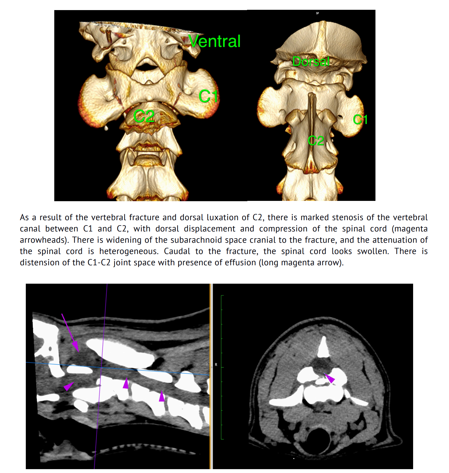
Image: Annotations and reconstructions support clear understanding of diagnostic imaging findings.

The Optics of Optimal Patient Care
For both the clinician and the patient, the benefits of annotated reports are clear and immediate:
- Improved Diagnostic Accuracy: Annotations ensure that imaging findings are explicitly identified. This precision leads to a more confident and accurate diagnosis, which is the first step toward effective treatment.
- Enhanced Communication: The visual nature of an annotated report makes it significantly easier to communicate complex medical findings to pet parents. Showing a client a clear image with an arrow pointing to a foreign body or a tumor helps them understand the diagnosis and the importance of the recommended treatment plan, fostering trust and compliance with the recommendations.
- Educational Value: Every annotated report is a learning opportunity. Veterinarians can review the marked images and use them to enhance their own diagnostic skills. Over time, this improves the practice’s overall capability to handle complex cases.
- Faster, More Confident Decisions: When faced with a critical case, quick well-informed decisions are needed. A quality assured report with annotated images provides clarity, enabling swift and confident action, which is vital for patient outcomes.

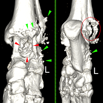
Image: Annotated images are an invaluable tool for explaining the rationale behind further diagnostic tests to reach a definitive diagnostic and support compliance with treatment recommendations.
Diagnostic interpretation:
BRAIN: The brain is abnormal in appearance with bilaterally symmetrical focal areas of homogenous T2 hyper-intensity within grey matter involving the caudal colliculi, vestibular nuclei, red nuclei and ventromedially in the thalamus. There are similar lesions in the midline of the ventral para-aqueductal grey matter and the cerebellar nodulus. There is no mass effect or atrophy associated with the lesions.
No diffusion abnormalities are visible and there are no hemorrhages on the T2* GRE images. On the Tiw images the lesions are iso-mildly hypointense. The post-contrast images show dense, moderate enhancement of the caudal colliculi and vestibular nuclei lesions. No other abnormal enhancement is visible. The conformation of the brain is normal with no gyral or sulcal abnormalities. The rest of the grey and white matter are normal in signal intensity with no signal alterations. The ventricular system is normal in size and shape. There is a normal size and shape to the pituitary gland. No cranial nerve abnormalities are visible. The extra-cranial structures are normal.

Note lesions in the para-aquaductal grey matter and thalamus Enhancement of the caudal colliculi lesions
Conclusions: Thiamine deficiency
Additional comments:
The presence of bilaterally symmetrical lesions is highly suggestive of a metabolic encephalopathy or toxicoses. The distribution of the lesions in this case is highly suggestive/pathognemonic for thiamine deficiency but definitive diagnosis would require measurement of thiamine levels. Enhancement of lesions in thiamine deficiency is seen sporadically and is not unusual.
The VET.CT Standard
As a leader in veterinary teleradiology, VET.CT has led the way in championing the use of annotated reports as a cornerstone of its service. We focus on the 6 C’s of good radiology reporting (Armas, 1998): Clarity, Correctness, Confidence, Concision, Completeness, and Consistency. Annotation enhances these factors and is also key to an effective 7th component of good radiology; communication - between the radiologist, clinician and client. Finally, we add the most important final ‘C’ of all - care. Our team genuinely care deeply about the patient, client and the veterinary team managing the vet-client-pet-relationship. We strive for good outcomes for all - not just the health and welfare of the patient, and the clinic’s relationship with their client, but helping the veterinary team to have a great day.
By sending diagnostic images to VET.CT, veterinarians gain access to a global network of over 150 board-certified radiologists. These experts meticulously review and provide detailed, visually rich reports, ensuring that every case receives the highest level of interpretation - a cornerstone of accurate and appropriate patient care.
VET.CT’s commitment to this practice ensures that veterinarians receive the support and reassurance they need, especially with complicated cases. It is a service built on our values, to bring our best, in delivering expert knowledge and support at the point of need, acting as a force for good in veterinary medicine.
By choosing to partner with a service that makes expert, annotated reports a standard, backed up by our unique clinical warranty, veterinarians are not just outsourcing a diagnostic step—they are elevating their practice. They are embracing improved diagnostic accuracy and streamlined communication, ultimately leading to better patient care and outcomes. The decision to send images VET.CT is a direct investment in the health and well-being of every animal you treat.
To find out more how VET.CT can support you and your team to give great patient care, contact us today to arrange a no obligation discovery call:

Ref: Armas RR, Qualities of a good radiology report. Am J Roentgenol. 1998; 170(4), https://doi.org/10.2214/ajr.170.4.9530077
Related article: The 8 C’s of Good Veterinary Diagnostic Imaging Reports
Explore more
Oct 23, 2025 2:48:04 PM
Master CT Imaging - Our expert-led webinars
What's on the agenda? - Introduction to CT Image Viewing & Quality Evaluation- Basics of CT...
Read more-1.jpg)
Oct 23, 2025 2:48:04 PM
Re-watch our latest veterinary webinar series
Our veterinary webinars were delivered by top global Board-certified radiologists and specialists...
Read more